≡ Menu

Task Stacking

If you work in an emergency room (ER), I am sure you have had a long journey to become more and more efficient. One way to be more efficient is to stack multiple tasks into the same cognitive process/execution. Here is what the beta version of ChatGPT has to say about it (https://openai.com):

Clearly, AI cannot yet educate an ER doctor on how to properly stack your tasks. So let me take a stab at it. ER docs love to learn from cases and examples, so I will present a few to illustrate my approach.

Here is an ideal scenario: you are signing out to the oncoming attending and you time a page to surgery to coincide with sign out of a patient. Once surgery calls you back, you say (while using body language to signal to the oncoming attending which patient you are talking about): Hey there, I have a 27 year old male who presented with 2 days of periumbilical pain, which then progressed to his right lower quadrant. He had tenderness to palpation in the right lower quadrant, so I got a ct scan, which showed non-perforated, acute appendicitis. His white count is 16, but isn’t septic. I gave a dose of zosyn and morphine for his pain. [hang up the phone, turn to the oncoming attending]. That’s Mr. XYZ in room 11.

At the same time of calling the consult, you have also given sign out to the oncoming attending. That probably saved one minute. If you can save 5 minutes a day, you will save about 20 hours a year. The more efficient you get, the more time you will save. Charting from home sucks, but you don’t have to do it if you get efficient.

Here is another example of multi task stacking: commuting to work on bicycle while listening to continuing medical education (CME) generating content. The stacked tasks include: exercise, saving money by not burning gas, continuing education, and commuting to work. The non-stacked alternative is driving to work in silence or listening to meaningless noise. In the first example, 4 things are effectively executed at the same time and contribute to productivity. In the latter example, only 1 thing is executed.

Finding tasks to stack is the challenging part. You may have found you already do some of this intuitively. But if you have a conscious approach to it, you can further maximize your efficiency. For example, unless you need to take a mindfulness break while you walk across the ED to gather suture supplies, you could also utilize that time to make a brief phone call, eg calling CT to let them know its time for the repeat head CT for the traumatic subarachnoid hemorrhage in spot 13.

In summary, task stacking is not a method, rather it is an approach. If you strive to make conscious decisions about your ED order of operations and optimize what tasks can be accomplished in parallel, you will find ways to stack more tasks. As a general rule of thumb, task stacking typically involves communicating to multiple parties at the same time or optimizing different tasks that use different parts of your brain/body. For me personally, utilizing this method has shifted the balance from finishing charts at home to finishing the vast majority of my charts on shift.

UPDATE 3.24.23

Here is GPT’s response to my post, when prompted:

GPT response to my post, when prompted.
GPT response 2
{ 0 comments }

Modified Rumack-Matthew Nomogram and Calculator

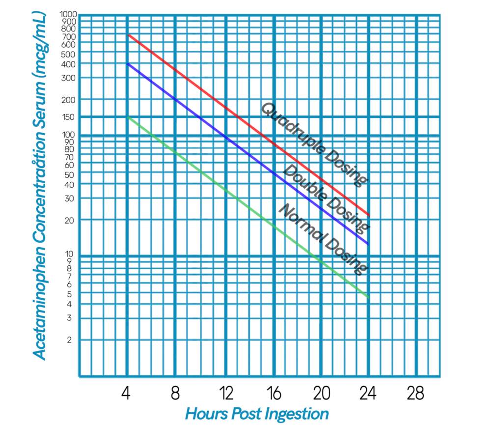
The purpose of the calculator is to eliminate visual estimation as a means for determination of need for treatment.

Normal dose NAC dosing compared to theoretical higher dosing based upon modified Rumack-Matthew nomogram

As per review of the literature, higher dose n-acetyl-cysteine (NAC) may be of benefit for patients with sub-massive and massive APAP overdose. More NAC means more glutathione, which means more capacity to detoxify the toxic metabolite of APAP, n-acetyl-para-benzoquinone imine (NAPQI). This modified nomogram gives theoretical recommendations for levels at which increased NAC doses may be considered.

Furthermore, 4-methylpyrazole (aka, fomepizole) is strong inhibitor of CYP2E1 and may decrease the rate at which NAPQI is formed. Therefore, 4-methylpyrazole may be considered as a safe adjunctive therapy in sub-massive and massive APAP overdoses.

This calculator and nomogram are intended for educational purposes only. This nomogram and calculator have not been clinically validated.

References:

Hazai E, Vereczkey L, Monostory K. Reduction of toxic metabolite formation of acetaminophen. Biochem Biophys Res Commun. 2002;291(4):1089-1094.

Hendrickson RG. What is the most appropriate dose of N-acetylcysteine after massive acetaminophen overdose? Clin Toxicol (Phila). 2019;57(8):686-691.

White SJ, Rumack BH. The acetaminophen toxicity equations: “solutions” for acetaminophen toxicity based on the rumack-matthew nomogram. Annals of Emergency Medicine. 2005;45(5):563-564.

{ 0 comments }

Rivastigmine for Antimuscarinic Delirium

In the setting of current drug shortage for the single manufacturer medication, IV physostigmine, we need to consider alternate options for routine treatment/diagnosis of antimuscarinic derlium. Interestingly, there is an oral formulation of physostigmine that has been reported to be available; however a quick query of pharmacies in a large metropolitan area (NYC) shows that it is unavailable as of 3/15/2022. Regardless of whether it existed in the past, oral agents are suboptimal for a delirious patient. The treatment of choice should be a tertiary amine acetylcholinesterase inhibitor (ACHe-i), given parenterally. There is only one other on the market: the rivastigmine patch.

Most likely a rivastigmine patch would be untenable for diagnosis of antimuscarinic delirium due to the pharmacokinetics. As per Micromedex®, the Tmax is anywhere between 8 and 16 hours, which is certainly much longer than IV formulation physostigmine, which seems to have peak effect with 5-10 minutes.

Kernebeek, et al recently reported that oral rivastigmine alleviated antimuscarinic delirium in a patient who had a confirmed procyclidine ingestion of over 300 tablets of 5mg. Apparently, they gave rivastigmine because IV physostigmine was not available (https://doi.org/10.1080/15563650.2020.1818768). It is important to note that this patient was intubated and subsequently extubated prior to administration of the PO rivastigmine. Although interesting, this single case report does little to add to the literature regarding an alternative to physostigmine. If the patient was well enough to be extubated due to resolving toxidrome, the benefit of PO rivastigmine is unclear.

From an ethical point of view, if there is a proven safe medication available, which we know is a theoretical antidote for antimuscarinic toxicity, there should be no qualm in recommending a rivastigmine patch, especially if dosed as per FDA regulation.

Other sources:

IBM micromedex

{ 0 comments }

Toxicology Flashcard set: Snakes and Spiders

{ 0 comments }

Titratable acid reserve

As medical toxicologists, we often get consulted on exposures to caustic agents. Caustic agents typically are at the extreme ranges of pH: eg, approaching 0 and 14 for acidic and alkali substances, respectively.

An important concept is the titratable acid reserve (TAR). Not all clinically significant caustic substances have extremes of pH. For example, glacial acetic acid is about a 17 Molar solution of acetic acid and water, which has pH of 2.4. However, an aqueous solution of hydrochloric acid with pH of 2.4 would only be a 0.004 Molar solution. So, despite having the same pH, glacial acetic acid would have 4250 times as many Moles of acid as the aforemention solution of HCl.

Hoffman, et al defined TAR as a the amount in mL of either 0.1M HCl or 0.1M NaOH required to bring 100 mL of solution to a pH of 8.0.

The general idea with TAR is that the more acid or base a particular xenobiotic takes to normalize a solution to a pH of 8.0, the more acid/base  living tissue exposed to that xenobiotic will have to neutralize. So, TAR is likely a better indicator of the caustic potential when compared to pH. The previously described solutions (glacial acetic acid and HCl both at pH 2.4) would likely have drastically different local tissue and systemic effects if ingested by a patient.  

In the 1989 Hoffman, et al paper, dissected dog esophaguses were exposed to a variety of caustic xenobiotics. Histologic damage was then assessed and referenced to each xenobiotic’s TAR. Although there is some overlap in TAR values 24-40, you can extrapolate that TAR less than 5 did not have any histologic damage; where as TAR greater than 40 universally had some demonstrable histologic damage. 

Examples of caustic agents with exceptionally high TAR include:

  • Phenol
  • Glacial Acetic Acid
  • Crystalline drain cleaner
  • 4% ammonia surface cleaner
  • Battery acid (eg, from lead based automotive battery)
  • Liquid toilet bowl cleaner

Examples of caustic agents with notably low TAR include: 

  • Over the counter bleach
  • Laundry and dish detergents 

So, utilizing TAR is likely a better way to guide clinical decision making regarding the decision to recommend admission for GI consult and possible endoscopy versus PO challenge and likely medical clearance. 

Further research could include update analysis of TAR in current and commonly available products available to the public. The resulting database of TAR’s could help further guide medical toxicologists decision making. 

References:

Hoffman RS, Howland MA, Kamerow HN, Goldfrank LR. Comparison of titratable acid/alkaline reserve and pH in potentially caustic household products. J Toxicol Clin Toxicol. 1989;27(4-5):241-246.

Goldfranks Toxicologic Emergencies 11e

{ 0 comments }

Base Excess: The forgotten lab?

In short, base excess (BE) represents the requisite amount of base required to titrate a blood sample to physiologic pH (7.35 to 7.45). Prior to the advent of rapidly available lactate concentration measurements, providers utilized BE as a surrogate marker for build up of organic acids, especially lactic acid.

BE has been largely supplanted by the ready availability of lactate testing, and thus is skipped over by most emergency medicine providers and toxicologists. However, there may be a reason to take a closer look at this laboratory value, especially when assessing poisoned patients.

How the test is performed:

  • Performed on standard blood gas analyzer
  • In the old days machine dilutes blood to 5 g/dL and standardizes pCO2 to 40 mm HG (note that the reason for dilution to 5 g/dL is that this is thought to account for fluids in the extracellular space); however, now this is done via a series of calculations performed by the machine. Our shop uses the Radiometer ABL800 Flex. See Derranged Physiology’s image of the equation here.
  • The sample is then titrated with either base or acid to a pH of 7.4
  • Output values represent the number of mmol/L of base required to titrate the sample to a pH of 7.4

Interpreting the test:

  • The more negative the result, the more base is required to normalize the sample; so, a more negative base deficit implicates that more acid is present in the sample
  • A positive base excess suggests a metabolic alkalosis is present

Potential benefits

  • Results are available rapidly, so if you are trending an acidosis you can have an answer in minutes versus having to wait about hour or more for the result of a BMP
  • Can be rapidly used in lieu of DeWinters formula because the blood gas result gives you pCO2 and BE. So, you can rapidly identify whether there is a respiratory acidosis or alkalosis AND a metabolic acidosis or alkalosis.

Examples:

  • A patient has a blood gas demonstrating a pH7.45, pCO2 of 20, BE of -20, and LA of 2.0. This patient has a respiratory alkalosis and a primary metabolic acidosis that is not likely attributable to a mild lactic acidosis. In a toxicologic setting, this patient’s blood gas is suggestive of salicylism.
  • A patient has a blood gas demonstrating a pH7.25, pCO2 of 60, BE of +4, and LA of 1.0. This patient has a respiratory acidosis with likely metabolic compensation, which suggests that the patient has a a chronic respiratory acidosis.
  • A patient has a blood gas demonstrating a pH7.51, pCO2 of 40, BE of +8, and LA of 1.5. This patient has a normal respiratory status and clearly has a primary metabolic alkalosis or may be on a sodium bicarbonate infusion.

Limitations:

  • Does not identify species of acids contributing to the acid-base status of the patient
  • Does not allow for deduction of mixed metabolic acidoses or combine metabolic acidosis/alkalosis; so, if the patient is compensating for a subacute acidosis, the BE may be higher.
  • Does not function for patients with a HGb of less than 5 gm/dL
  • Should be correlated with concomitant BMP’s to calculate the AG, other objective data (including vital signs), as well as the clinical history.

References:

Berend K. Diagnostic use of base excess in acid-base disorders. N Engl J Med. 2018;378(15):1419-1428.

https://derangedphysiology.com/main/cicm-primary-exam/required-reading/acid-base-physiology/Chapter%20603/actual-base-excess

{ 0 comments }

Tech Security

I’ve included a PDF of a document for securing your personal devices. Needless to say, it is quite obvious the majority of health care professionals do none of this. However, I highly recommend enhancing your focus on personal digital security. For example, if your laptop is stolen and the hard drive is not encrypted, all your cookies, emails, and login information can be accessed by a skilled criminal. See my guide, attached as a PDF to take your digital security to the industry standard.

The purpose of this document is to provide a guide for improving security of personal devices. This guide currently only supports devices within Apple Ecosystem. Encryption and security of devices is critically important in the event of loss of a personal device. Whether it is stolen or mistakenly lost and unable to be recovered, an unsecured device risks loss of data to unauthorized entities. You would be surprised the amount of data stored on your computer or phone that you didn’t know was there. All your saved logins, email attachments in temporary folders, credit card information, work related data, cookies from your browsing, etc…. the list goes on and on. 

How to encrypt your MacOS personal computer: 

  1. Make sure your computer password is distinct from any other password you use at your institution. and equally as robust. 
  2. Click the Apple symbol () in the upper left hand computer. 
  3. Go to “System Preferences”
  4. Click “Security & Privacy”
  5. Click “Turn On FileVault” (perform this overnight, as it may take some time)
  1. You can set a recovery key in your iCloud. This should have a password distinct from other passwords and be as robust, with a variety of alphanumeric symbols.
  2. Now as proof of encryption, click the Apple symbol () in the upper left hand corner, click the “About This Mac”. Next place the “Security & Privacy” window next to the information window.  Take a screen shot (command, shift, option 4) and crop the screen area utilizing the target symbol and your cursor.

  1. Open Preview app. Press command+n. Save this and back it up online, preferably in a secured email. In the event of data breach, this can be used as proof any PHI that may be on the personal computer is secure. 
  2. You should also enable FileVault for any usb backup drives or usb peripheral drives. 

Enable firmware password on your Apple computer: 

  1. Enabling a firmware password on your computer will prevent users from being able to boot your computer from an external drive, unless they know the firmware password. It may seem like overkill, but this is the final step to complete data protection
  2. Follow this link to the Apple website for a detailed guide: https://support.apple.com/en-us/HT204455 

How to encrypt your iPhone: 

  1. Requiring a passcode to open your iPhone now automatically encrypts your iPhone. If you chose to use or organization’s email/VPN on your iPhone, you likely will be required to enter an alphanumeric passcode which will further enhance security. 

How to further secure your iPhone:

  1. Open “Settings”
  2. Open “Face ID & Passcode”
  3. Enable Erase Data after 10 failed attempts. This will further protect possible breach of PHI.   
{ 0 comments }

Catecholamine Metabolic Pathway

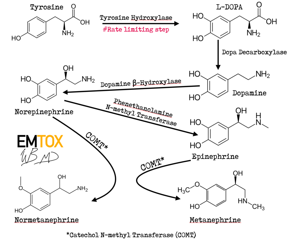
So during my studies, I realized that there aren’t many good/clear images of the catecholamine metabolic pathway that are easily searchable via Google images. So, I made my own figure. Everything is adapted from the public domain, but crisped up with some 2020 technology.

The important takeaway here is that tyrosine hydroxylase is the rate limiting step for catecholamine synthesis (see this 1964 article by Nagatsu, et al; PMID: 14216443). Keep in mind the pH optimum for tyrosine hydroxylase was determined to be in around 6, but the experimental procedure that measured radio-labeled metabolites was performed at pH8.5. Interestingly, activity of tyrosine hydroxylase is dependent on the tetrohydrofolate derivative: tetrahydropteridine. So, the rate limiting step of catecholamine synthesis appears to be folate dependent….

A brief pubmed search suggests that no randomized clinical trial has been performed on folate supplementation in shock states. Why are sepsis researchers focusing on “skin vitamins” in the past?

{ 0 comments }